| Overview of field and research projects |
The Dept. of Medical Technologies and Sciences focuses on three main areas of study: (1) computer-aided diagnosis (CAD), (2) development of diagnostic imaging using radioactive isotopes, and (3) optimization of radiation dosage in medical CT examinations.
- (1)In the laboratory of Image Information Sciences, research and development of CAD (Fig.1) is carried out. CAD is defined as the use of the results of computer quantitative analysis of affected areas of patients in medical images as a second medical opinion for diagnostic purposes. It has a wide range of applications, including diagnostic imaging, radiology, and ultrasound analysis (Fig.2). Further development of CAD would increase accuracy of medical image diagnosis. Receiver operating characteristic (ROC) analysis is used to evaluate the accuracy of diagnoses via CAD for use in clinical applications. Future developments in this field will lead to the application of medical image diagnosis technology for quantitative diagnostic therapy.(http://www.img.hs.kumamoto-u.ac.jp/)
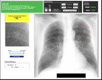 Figure 1 : CAD output for the classification of lung nodules on chest image. 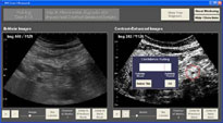 Figure 2 : Computer interface for the detection of liver metastases. - (2)In the laboratory of Molecular Function Research, new internal imaging diagnostic methods (SPECT, PET, etc.) are being developed through the synthesis of molecules to react to specific diseases, labeling of useful RI (131I, 125I, 18F, 11C, etc.), and intravenous injection of labeled compounds. Notable developments include clinical applications of cancer diagnosis with 18FAMT (Fig.3) and Parkinson's disease diagnosis with 18FmT (Fig.4) using functional imaging through PET analysis. Currently, we are conducting experiments for clinical diagnosis of amyloidosis by improving the usefulness of IP imaging (Fig.5) of 125I-EMSB in normal rat brains as part of our research on diagnostic imaging of amyloidosis and Alzheimer's disease, which causes dementia (18F-EMSB, 125I-EMSB).
(http://www.molimaging.hs.kumamoto-u.ac.jp/)
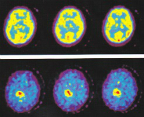 Figure 3 : Upper : 18FDG, Lower: 18FAMT in same astrocytoma patient. 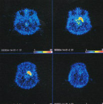 Figure 4 : PET slices of 18FmT including the striatum in a Parkinson Monkey. 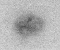 Figure 5 : IP imaging of 125I-EMSB in a rat sagittal brain - (3)In the laboratory of Tomographic Imaging, researchers aim to develop more accurate clinical diagnoses through the optimization of CT X-ray dosage without compromising image quality by using image reconstruction and image processing technologies. We are conducting research on the optimization of dosage and image quality using cardiac CT:FBP and IR methods, low voltage technology in prospective and retrospective cardiac CT, evaluation of cardiovascular stents with IR methods and high-resolution reconstruction functions, reduction of the dosage of radioactivity and contrast medium in pediatric CT:CNR methods, optimization of the dosage of radioactivity in temporal bone CT, and virtual liver phantoms present in low signal and high signal abdominal CT.
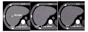 Figure 6 : Virtual liver phantom with hypo nodule acquired at 100, 50, and 25% x-ray doses
|
|---|